44 heart structure and labels
Heart Diagram with Labels and Detailed Explanation - BYJUS Well-Labelled Diagram of Heart. The heart is made up of four chambers: The upper two chambers of the heart are called auricles. The lower two chambers of the heart are called ventricles. The heart wall is made up of three layers: The outer layer of the heart wall is called epicardium. The middle layer of the heart wall is called myocardium. How to Draw the Internal Structure of the Heart (with ... Step 1, To find a good diagram, go to Google Images, and type in "The Internal Structure of the Human Heart". Find an image that displays the entire heart, and click on it to enlarge it.Step 2, Find a piece of paper and something to draw with. Start with the pulmonary veins. They will be to the lower left of the Aorta. There are two of them. Draw the top vein slightly smaller than the bottom vein.Step 3, Below the pulmonary veins, and slightly to the right, begin sketching the bottom of the ...
Structure and Function of the Heart - ScienceGeek.net Structure and Function of the Heart. The Human Heart. Refer to this diagram when answering the questions on the right. ... The ascending vena cava is a major vein that returns deoxygenated blood to the heart from the lower part of the body. In this diagram, it is labeled.

Heart structure and labels
Structure Of The Heart | A-Level Biology Revision Notes The heart is a hollow muscular organ that lies in the middle of the chest cavity. It is enclosed in the pericardium, which protects the heart and facilitates its pumping action. The heart is divided into four chambers: The two atria (auricles): these are the upper two chambers. The structure of the heart - Structure and function of the ... It is located in the middle of the chest and slightly towards the left. The heart is a large muscular pump and is divided into two halves - the right-hand side and the left-hand side. The... Layers of the heart: Epicardium, myocardium, endocardium ... The heart is a muscular organ found in the middle mediastinum that pumps blood throughout the body. It is housed in the pericardial sac, which protects it and assists with its mechanics. Recalling from the heart anatomy, it has two atria and two ventricles that make up elements and important steps for the heart cycle.
Heart structure and labels. heart | Structure, Function, Diagram, Anatomy, & Facts ... A thin layer of tissue, the pericardium, covers the outside, and another layer, the endocardium, lines the inside. The heart cavity is divided down the middle into a right and a left heart, which in turn are subdivided into two chambers. The upper chamber is called an atrium (or auricle), and the lower chamber is called a ventricle. Heart Anatomy | Anatomy and Physiology ventricle: one of the primary pumping chambers of the heart located in the lower portion of the heart; the left ventricle is the major pumping chamber on the lower left side of the heart that ejects blood into the systemic circuit via the aorta and receives blood from the left atrium; the right ventricle is the major pumping chamber on the lower right side of the heart that ejects blood into the pulmonary circuit via the pulmonary trunk and receives blood from the right atrium Labelling the heart — Science Learning Hub Labelling the heart — Science Learning Hub Labelling the heart Add to collection The heart is a muscular organ that pumps blood through the blood vessels of the circulatory system. Blood transports oxygen and nutrients to the body. It is also involved in the removal of metabolic wastes. Topics Concepts Citizen science Teacher PLD Glossary Sign in Heart Anatomy Labeling Game This is an online quiz called Heart Anatomy Labeling Game. There is a printable worksheet available for download here so you can take the quiz with pen and paper. Your Skills & Rank. Total Points. 0. Get started! Today's Rank--0. Today 's Points. One of us! Game Points. 19. You need to get 100% to score the 19 points available.
Structure of the Heart | Biology for Majors II The heart is composed of three layers; the epicardium, the myocardium, and the endocardium, illustrated in Figure 1. The inner wall of the heart has a lining called the endocardium.The myocardium consists of the heart muscle cells that make up the middle layer and the bulk of the heart wall. The outer layer of cells is called the epicardium, of which the second layer is a membranous layered ... The Anatomy of the Heart, Its Structures, and Functions The Cardiac cycle is the sequence of events that occurs when the heart beats. Below are the two phases of the cardiac cycle: Diastole phase: The heart ventricles are relaxed and the heart fills with blood. Systole phase: The ventricles contract and pump blood to the arteries. Valves PDF Anatomy of Heart Labeled and Unlabeled Images ascending aorta pulmonary valve opening of superior vena cava right atrium fossa ovalis tricuspid valve right ventricle trabeculae carneae (a) anterior dissection of the heart 2019 pearson education, inc, pu monary trunk openings of left pulmonary veins left atrium aortic valve mitral (bicuspid) va ve chordae tendineae papillary muscle left … Heart Anatomy: Labeled Diagram, Structures, Blood Flow ... By the end of this post, you will have a strong understanding of the main cardiac structures including: Atria. Ventricles. Tricuspid Valve. Mitral Valve. Pulmonary Valve. Aortic Valve. Superior and Inferior Vena Cava. Pulmonary Arteries and Veins. Aorta. You will also be provided with numerous memory tricks to help you remember the different structures of the heart!
Heart Anatomy - GetBodySmart Heart Anatomy. An online interactive study guide to tutorials and quizzes on the anatomy and physiology of the heart, using interactive animations and diagrams. Looking for free labeling diagrams? PDF HEART - STRUCTURE - BiologyMad HEART - STRUCTURE • 4 sections Left atrium Right atrium Left ventricle Right ventricle • heart ry artery Pulmonary vein EAS the blood from he left hand side has to be pumped all around the body. • 2 lo heart Atrioventricular valves - between the atrium and the ventricles Semi-lunar valves - in the pulmonary artery and the aorta Heart anatomy: Structure, valves, coronary vessels | Kenhub Inside, the heart is divided into four heart chambers: two atria (right and left) and two ventricles (right and left). Heart Labeling Quiz: How Much You Know About Heart ... Here is a Heart labeling quiz for you. The human heart is a vital organ for every human. The more healthy your heart is, the longer the chances you have of surviving, so you better take care of it. Take the following quiz to know how much you know about your heart. Questions and Answers 1. What is #1? 2. What is #2? 3. What is #3? 4. What is #4?
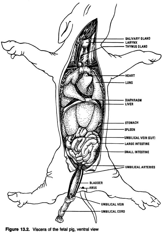
Anatomical Drawings of a Fetal Pig
Heart Blood Flow | Simple Anatomy Diagram, Cardiac ... One of the first things you will notice if you look at the 12 steps is the pattern between the right and left side of the heart is similar. Step 1 and 6 involve a blood vessel, which makes sense as this is how blood enters and exits that side of the heart. Steps 2-5 involve a chamber, valve, chamber, and valve.

China U. S Trade War Heading To Economic Collapse : heading,News, breakingnews, globalnews ...
Heart - Wikipedia The wall of the heart is made up of three layers: epicardium, myocardium, and endocardium. The heart pumps blood with a rhythm determined by a group of pacemaker cells in the sinoatrial node. These generate a current that causes the heart to contract, traveling through the atrioventricular node and along the conduction system of the heart.

Histology Drawings: January 2014
Human Heart - Anatomy, Functions and Facts about Heart One of the very first structures which can be observed when the external structure of the heart is viewed is the pericardium. Pericardium The human heart is situated to the left of the chest and is enclosed within a fluid-filled cavity described as the pericardial cavity.

Pin by Daffodilcooper on BSC2086 | Heart model, Anatomy models labeled, Cardiac anatomy
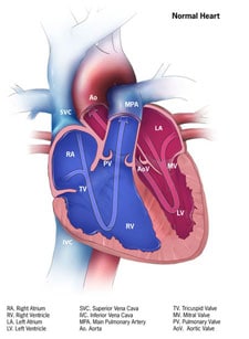
Label the heart - Science Learning Hub Labels. Description. Vena cava. Carries deoxygenated blood from the body to the heart. Semilunar valve. Flaps that prevent backflow of blood. Left atrium. Receives oxygenated blood from the lungs. Left ventricle. Region of the heart that pumps oxygenated blood to the body. Pulmonary artery. Carries deoxygenated blood to the lungs. Right ventricle
Drawing Of The Brain With Labels | Free download on ClipArtMag
Structure of the Heart - The Franklin Institute Structure of the Heart Although most people know that the human heart doesn't bear much resemblance to a heart drawn on a Valentine's Day card, the image can still be a useful way to learn and remember the parts of the heart.
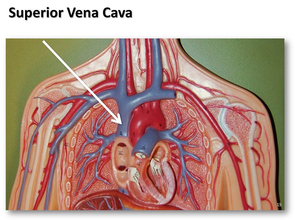
Superior vena cava - The Anatomy of the Veins Visual Guide… | Flickr
A Labeled Diagram of the Human Heart You Really Need to ... The human heart, comprises four chambers: right atrium, left atrium, right ventricle and left ventricle. The two upper chambers are called the left and the right atria, and the two lower chambers are known as the left and the right ventricles. The two atria and ventricles are separated from each other by a muscle wall called 'septum'.
How the Heart Works | Congenital Heart Defects | NCBDDD | CDC
Structure of the Heart | SEER Training Chambers of the Heart. The internal cavity of the heart is divided into four chambers: Right atrium; Right ventricle; Left atrium; Left ventricle; The two atria are thin-walled chambers that receive blood from the veins. The two ventricles are thick-walled chambers that forcefully pump blood out of the heart. Differences in thickness of the heart chamber walls are due to variations in the amount of myocardium present, which reflects the amount of force each chamber is required to generate.

Ask Thucydides! (“The Baker Street Irregulars’ ‘Thucydides’ whose Archival Series has set the ...
Anatomy of the heart and blood vessels | Patient The heart is a muscular pump that pushes blood through blood vessels around the body. The heart beats continuously, pumping the equivalent of more than 14,000 litres of blood every day through five main types of blood vessels: arteries, arterioles, capillaries, venules and veins.
Post a Comment for "44 heart structure and labels"