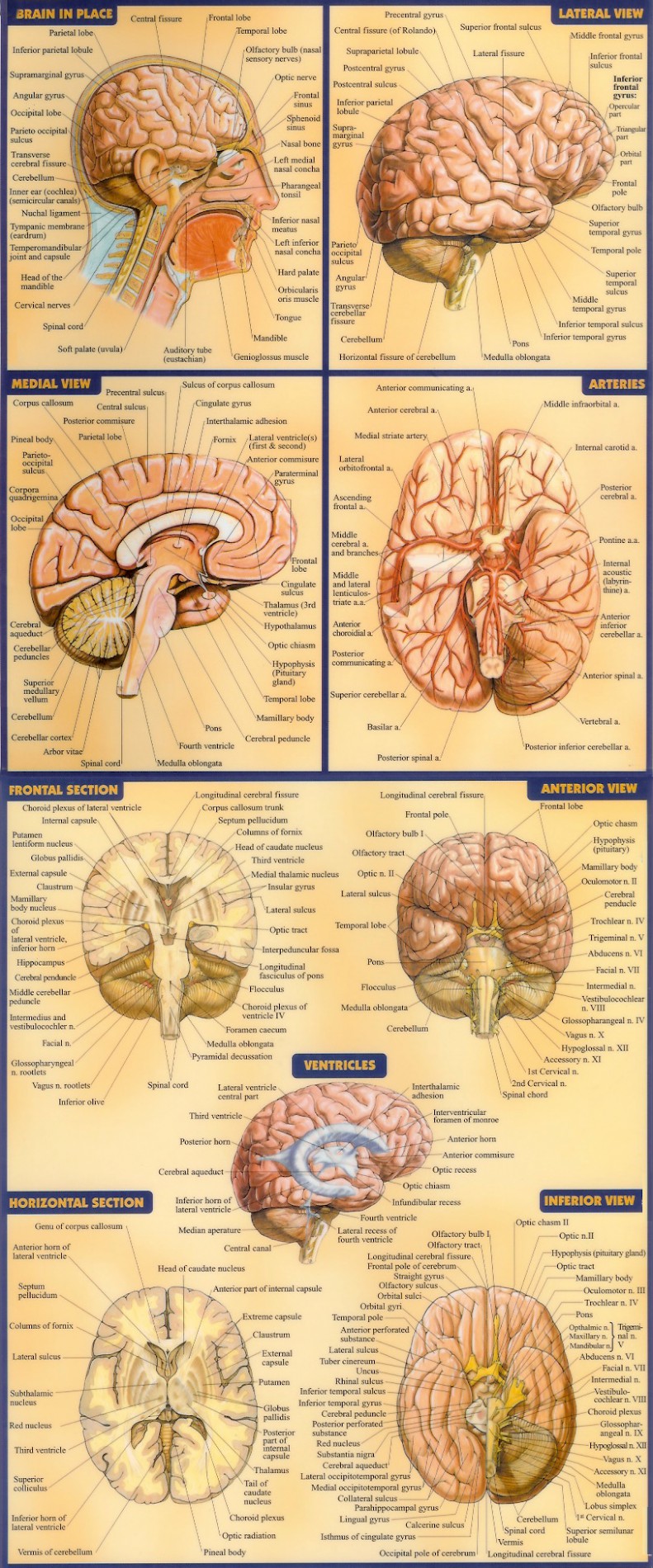
39 the brain diagram with labels
Artificial Neural Network | Brilliant Math & Science Wiki The human brain is primarily comprised of neurons, small cells that learn to fire electrical and chemical signals based on some function.There are on the order of 1 0 11 10^{11} 1 0 1 1 neurons in the human brain, about 15 15 1 5 times the total number of people in the world. Each neuron is, on average, connected to 10000 10000 1 0 0 0 0 other neurons, so that there are a total of 1 0 15 10 ... Structure of the Brain and Their Functions - New Health Advisor It takes up 60% of brain matter. Numbers- The brain is made up of 75% water. It has 60% fat thus making it the fattest organ in the body. It has at least some 100,000 miles of blood vessels in the brain. The brain generates between 10 -23 watts of energy whilst you are awake and this is sufficient to light up a bulb.
Cross sectional anatomy | Kenhub Cross section through the thalamus: Diagram Orienting yourself within such a cross section is easy. The star of the show (brain) is easily recognizable because it appears highly convoluted, full of ridges (gyri) and indentations (sulci).The paired thalami appear as two circular masses in the midline, forming the walls of the third ventricle.The neurocranium appears as a meshwork (trabecular ...
The brain diagram with labels
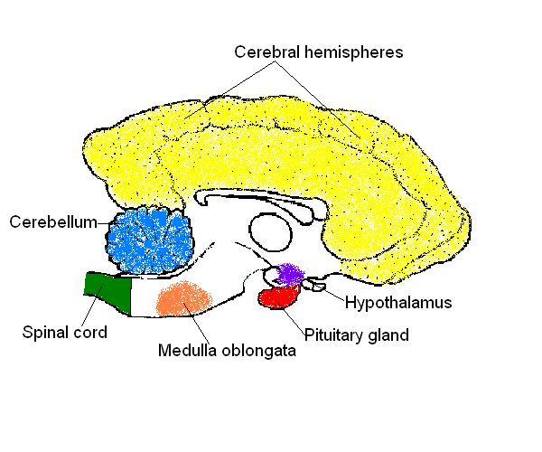
wikieducator.org › Nervous_System_Worksheet_AnswersNervous System Worksheet Answers - WikiEducator Jan 14, 2008 · 8. The diagram below shows a section of a dog’s brain. Add the labels in the list below and, if you like, colour in the diagram as suggested. Cerebellum - blue; Spinal cord - green; Medulla oblongata - orange; Hypothalamus - purple; Pituitary gland - red; Cerebral hemispheres – yellow. 9. Match the descriptions below with the terms in the list. ADHD Aetiology | ADHD Institute Structural abnormalities that have been observed in children, adolescents and adults with ADHD versus individuals without ADHD include: lower grey matter density 1-3; white matter abnormalities 4,5; reduced total brain volume and volume of some brain structures 1,6-8,28; delayed cortical maturation in children and adolescents 9-11; and reduced cortical thickness in adults. 1,12 Action goals and the praxis network: an fMRI study - Brain Structure ... The praxis representation network (PRN) of the left cerebral hemisphere is typically linked to the control of functional interactions with familiar tools. Surprisingly, little is known about the PRN engagement in planning and execution of tool-directed actions motivated by non-functional but purposeful action goals. Here we used functional neuroimaging to perform both univariate and multi ...
The brain diagram with labels. Science vs. the "Gay Gene" - True.Origin In a similar manner, the brain was once considered to be rigid, like Ball ® jars used for canning—but we now know the brain is "plastic" and flexible, and able to reorganize itself. Research has shown that the brain is able to remodel its connections and grow larger, according to the specific areas that are most frequently utilized. 4 Types of Parenting Styles and Their Effects On The Child Different styles of parenting can lead to different child development and child outcomes. Based on extensive observation, interviews, and analyses, Baumrind initially identified these three parenting styles: authoritative parenting, authoritarian parenting, and permissive parenting 1 . credit: wikipedia.com. Raptor Vision - Oxford Research Encyclopedia of Neuroscience The axons of RGCs reach the brain via the optic nerve, after a complete nerve crossing in birds, and deliver information to three parallel visual systems: tectofugal pathway, thalamofugal pathway, and accessory optic system (reviewed in Güntürkün, 2000 or Wylie, Gutiérrez-Ibáñez, & Iwaniuk, 2015). The majority of RGCs in birds contribute ... byjus.com › biology › liver-diagramLiver Diagram with Detailed Illustrations and Clear Labels Liver Diagram The liver is one of the most important organs in the human body. Anatomically, the liver is a meaty organ that consists of two large sections called the right and the left lobe.
Diagram of Human Heart and Blood Circulation in It Four Chambers of the Heart and Blood Circulation. The shape of the human heart is like an upside-down pear, weighing between 7-15 ounces, and is little larger than the size of the fist. It is located between the lungs, in the middle of the chest, behind and slightly to the left of the breast bone. The heart, one of the most significant organs ... Ethmoid bone: Anatomy, borders and development | Kenhub Relations. The ethmoid bone is a spongy, irregular bone of the skull. It is located anteriorly in the cranial base and contributes to the formation of the medial walls of the orbit, the nasal septum, and the roof and lateral walls of the nasal cavity.. Because of its central location within the skull the ethmoid bone comes in contact with 13 skull bones (e.g. frontal bone, sphenoid bone ... The Psychopathic Brain: Is It Different from a Normal Brain? The psychopathic brain has been an interest of study for decades due to the fact that psychopaths represent such a small segment of society and yet commit a highly disproportionate amount of criminal acts (like these famous psychopaths and psychopath killers).And with more-and-more psychopaths having magnetic resonance imaging (MRI) scans or functional MRI (fMRI) scans, some correlates have ... Brain - Wikipedia A brain is an organ that serves as the center of the nervous system in all vertebrate and most invertebrate animals. It is located in the head, usually close to the sensory organs for senses such as vision.It is the most complex organ in a vertebrate's body. In a human, the cerebral cortex contains approximately 14-16 billion neurons, and the estimated number of neurons in the cerebellum is ...
The Cerebrospinal Venous System: Anatomy, Physiology, and ... - Medscape Caudally, the CSVS freely communicates with the sacral and pelvic veins and the prostatic venous plexus. The CSVS constitutes a unique, large-capacity, valveless venous network in which flow is bidirectional. The CSVS plays important roles in the regulation of intracranial pressure with changes in posture, and in venous outflow from the brain. Head Truth: Don't Be Fooled: Well-Trained Chatbots Aren't Minds The Guy with the Smallest Brain Had the Highest IQ; He Had Half a Brain and Above Normal Intelligence; The Truth About Neurons and Synapses; A Diagram of Explanatory Dysfunction in Academia; The Brain Shows No Sign of Working Harder During Thinking or Recall; More Evidence of High Mental Function Despite Large Brain Damage Anatomy Project - Sheridan College Neck. · Connecting the shaft and head of the femur. · Projects superior and medial from the shaft to the head. · In addition to projecting superior and medial from the shaft of the femur, the neck also projects somewhat anterior. · The amount of forward projection is extremely variable, but on an average is from 12° to 14°. Human brain - Wikipedia The human brain is the central organ of the human nervous system, and with the spinal cord makes up the central nervous system.The brain consists of the cerebrum, the brainstem and the cerebellum.It controls most of the activities of the body, processing, integrating, and coordinating the information it receives from the sense organs, and making decisions as to the instructions sent to the ...
developingchild.harvard.edu › resources › the-brainThe Brain Circuits Underlying Motivation: An Interactive Graphic The brain systems that govern motivation are built over time, starting in the earliest years of development. These intricate neural circuits and structures are shaped by interactions between the experiences we have and the genes we are born with, which together influence both how our motivation systems develop and how they function later in life.
brainly.com › question › 11404375The diagram shows the electric field around two charged ... In the given diagram,the filed lines for W is towards W itself.The same is also in case of X. Hence both the charges must be negative in nature. Hence the correct answer to the question will be B i.e W negative and X: NEGATIVE.
Ocular Adnexa: Definition & Anatomy - Study.com The term for accessory structures of the eye is ocular adnexa. 'Ocular' refers to eyes and 'adnexa' is a Latin term meaning 'fasten to' and in this case refers to accessory structures attached to ...
A novel microRNA, novel-m009C, regulates methamphetamine rewarding ... Specific riboprobe for novel-m009C was fluorescently labeled (5'CY3 and 3'CY3, GenePharma, Shanghai) and used at a final labeled nucleotide concentration of 1.3 μM/l.
Gram Stain Technique - Amrita Vishwa Vidyapeetham Label one side of the glass slide with 1. Your initials 2. The date; While flaming the inoculation loop be sure that each segment of metal glows orange/red-hot before you move the next segment into the flame. Once you have flamed your loop, do not lay it down, blow on it, touch it with your fingers, or touch it to any surface other than your ...
Free Brain Diagram, Download Free Brain Diagram png images, Free ClipArts on Clipart Library
› photos › diagram-of-bodyDiagram Of Body Organs Female Pics Stock Photos, Pictures ... Human internal organs Internal organs in woman and man body. Brain, stomach, heart, kidney, medical icon in female and male silhouette. Digestive, respiratory, cardiovascular systems. Anatomy poster vector illustration. diagram of body organs female pics stock illustrations
What is the pathophysiology of borderline personality ... - Medscape Reports also indicate that adults with BPD have increased impulsivity, cognitive inflexibility, and poor self-monitoring and perseveration, which may be indicators of frontal lobe dysfunction. The ...
Intellectual Disability: Causes and Characteristics - HealthyPlace Intellectual disability causes children with the condition to take longer than typical children to sit, crawl, walk, speak, and take care of their personal needs. They have trouble learning at the same rate as other kids in school. Impaired children experience considerable challenges in two primary areas: intellectual functioning and adaptive ...

Drag each label to the correct location on the diagram. label the parts of the brain - Brainly.com
Whole-Brain Wiring Diagram of Oxytocin System in Adult Mice Oxytocin (Oxt) neurons regulate diverse physiological responses via direct connections with different neural circuits. However, the lack of comprehensive input-output wiring diagrams of Oxt neurons and their quantitative relationship with Oxt receptor (Oxtr) expression presents challenges to understanding circuit-specific Oxt functions. Here, we establish a whole-brain distribution and ...
Brain Structures and Their Functions | MD-Health.com Brain Stem. All basic life functions originate in the brain stem, including heartbeat, blood pressure and breathing. In humans, this area contains the medulla, midbrain and pons. This is commonly referred to as the simplest part of the brain, as most creatures on the evolutionary scale have some form of brain creation that resembles the brain ...
How the Brain Changes During Depression Treatment - Satrn News University of British Columbia researchers have mapped what occurs in the brain when a person gets the depression treatment known as repetitive transcranial magnetic stimulation.A new study maps how the brain changes throughout depression treatmentResearchers have for the first time shown what occurs in the brain during repetitive transcranial magnetic stimulation, a treatment for depression…
Accessory nerve | Radiology Reference Article | Radiopaedia.org The cranial part (accessory portion) is the smaller of the two. Its fibers arise from the cells of the nucleus ambiguus and emerge as four or five delicate rootlets from the side of the medulla oblongata, below the roots of the vagus nerve. It runs laterally to the jugular foramen, where it interchanges fibers with the spinal portion or becomes ...
Properties of Rectangles | Brilliant Math & Science Wiki In the diagram above, each circle is in contact two other circles and at least one side of the rectangle. The radii are perpendicular to the sides of the rectangle as shown. Find the area of the shaded portion in cm 2 \text{cm}^2 cm 2 to the nearest whole number. All dimensions are in cm. Use π ≈ 3.14159 \pi \approx 3.14159 π ≈ 3. 1 4 1 5 9.
Positions and Functions of the Four Brain Lobes - MD-Health.com The occipital lobe, the smallest of the four lobes of the brain, is located near the posterior region of the cerebral cortex, near the back of the skull. The occipital lobe is the primary visual processing center of the brain. Here are some other functions of the occipital lobe: Visual-spatial processing. Movement and color recognition.
en.wikipedia.org › wiki › File:Human_skeleton_frontFile:Human skeleton front en.svg - Wikipedia Restructured the image internals by adding layers, changing groupings, and adding meaningful ids and labels so that the image is easier to manipulate programmatically. Also made the labels text elements and gave them ids (it might be possible to generate : 10:17, 1 October 2007: 436 × 842 (764 KB) LadyofHats: some changes asked in FP discussion
Using Basic Plotting Functions - Video - MATLAB - MathWorks This includes adding titles, axes labels, and legends, and editing a plot's lines and markers in shape, style, and color. For more information on plotting, you can use help and documentation right from within MATLAB. Related Products. MATLAB; Bridging Wireless Communications Design and Testing with MATLAB. Read white paper.



Post a Comment for "39 the brain diagram with labels"