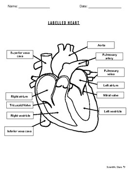
43 heart structure with labels
Diagrams, quizzes and worksheets of the heart | Kenhub Worksheet showing unlabelled heart diagrams. Using our unlabeled heart diagrams, you can challenge yourself to identify the individual parts of the heart as indicated by the arrows and fill-in-the-blank spaces. This exercise will help you to identify your weak spots, so you'll know which heart structures you need to spend more time studying ... Simple heart diagram | Simple heart diagram labeled - Pinterest Sep 16, 2020 - Simple heart diagram | Simple heart diagram labeled | Human heart diagram.We provide you a simple heart diagram to draw and learn. Simple heart diagram labeled with accurate labels. Most frequent question in exam to draw human heart diagram with labels. You can learn diagram of heart with labels and easy simple heart anatomy with heart structure.
Heart Diagram with Labels and Detailed Explanation The heart is located under the ribcage, between the lungs and above the diaphragm. It weighs about 10.5 ounces and is cone shaped in structure. It consists of the following parts: Heart Detailed Diagram Heart - Chambers There are four chambers of the heart . The upper two chambers are the auricles and the lower two are called ventricles.

Heart structure with labels
Label the Heart Diagram | Quizlet Label the Heart STUDY Learn Write Test PLAY Match Created by bluesas9 Terms in this set (15) Superior Vena Cava ... Right Ventricle ... Left Atrium ... Atrioventricular/Tricuspid Valve ... Atrioventricular/Mitral Valve ... Septum ... Right Atrium ... Semi-lunar Valves ... Left Pulmonary Veins ... Right Pulmonary Veins ... Left Pulmonary Arteries Heart Labeling Quiz: How Much You Know About Heart Labeling? Here is a Heart labeling quiz for you. The human heart is a vital organ for every human. The more healthy your heart is, the longer the chances you have of surviving, so you better take care of it. Take the following quiz to know how much you know about your heart. Questions and Answers 1. What is #1? 2. What is #2? 3. What is #3? 4. What is #4? Label the heart — Science Learning Hub In this interactive, you can label parts of the human heart. Drag and drop the text labels onto the boxes next to the diagram. Selecting or hovering over a box will highlight each area in the diagram. Right ventricle Right atrium Left atrium Pulmonary artery Left ventricle Pulmonary vein Semilunar valve Vena cava Aorta Download Exercise Tweet
Heart structure with labels. Human Heart (Anatomy): Diagram, Function, Chambers, Location in ... - WebMD The heart is a muscular organ about the size of a fist, located just behind and slightly left of the breastbone. The heart pumps blood through the network of arteries and veins called the... Label the HEART | Circulatory System Quiz - Quizizz 24 Questions Show answers Question 1 60 seconds Q. What is number two pointing at in the heart diagram? answer choices Right Atrium Right Ventricle Left Atrium Left Ventricle Question 2 60 seconds Q. What is number one pointing at in the heart diagram? answer choices Right Ventricle Right Pulmonary Vein Superior Franklin Inferior Vena Cava The structure of the heart - Structure and function of the heart ... Each side of the heart consists of an atrium and a ventricle which are two connected chambers. The atria (plural of atrium) are where the blood collects when it enters the heart. The ventricles... The Anatomy of the Heart, Its Structures, and Functions The heart is the organ that helps supply blood and oxygen to all parts of the body. It is divided by a partition (or septum) into two halves. The halves are, in turn, divided into four chambers. The heart is situated within the chest cavity and surrounded by a fluid-filled sac called the pericardium. This amazing muscle produces electrical ...
Structure and Function of the Heart - Medical News The heart is a muscle whose working mechanism is made possible by the many parts that operate together. The organ is divided into several chambers that take in and distribute oxygen-poor or oxygen ... heart | Structure, Function, Diagram, Anatomy, & Facts | Britannica heart, organ that serves as a pump to circulate the blood. It may be a straight tube, as in spiders and annelid worms, or a somewhat more elaborate structure with one or more receiving chambers (atria) and a main pumping chamber (ventricle), as in mollusks. In fishes the heart is a folded tube, with three or four enlarged areas that correspond to the chambers in the mammalian heart. In animals ... heart structure diagram labeled heart structure diagram labeled Sea Urchin- Enchanted Learning Software we have 9 Pics about Sea Urchin- Enchanted Learning Software like Diagram of Heart Blood Flow for Cardiac Nursing Students - NCLEX Quiz, Label parts of the heart interactive and downloadable worksheet. You and also Smart Label Parts of a Heart Exercise - Knomadix. Here you go: 20 POINTS AVAILABLE Identify the structures of the heart. Label A Label ... Septum is the tissue that separates left and right ventricles while aorta is the main artery which carry blood from heart to tissues. Thus, label A is pulmonary valve, label B is tricuspid valve, label C is septum, label D is bicuspid valve, label E is aortic valve and label F is aorta. For more information about structure of heart, visit:
Heart Anatomy: Labeled Diagram, Structures, Function, and Blood Flow There are 4 chambers, labeled 1-4 on the diagram below. To help simplify things, we can convert the heart into a square. We will then divide that square into 4 different boxes which will represent the 4 chambers of the heart. The boxes are numbered to correlate with the labeled chambers on the cartoon diagram. Human Heart: Label the diagram 1 - Liveworksheets Human Heart: Label the diagram 1 worksheet. Live worksheets > English. Human Heart: Label the diagram 1. Study the figure carefully.Label the 10 parts of the human heart A-J. ID: 1781041. Language: English. School subject: Biology. Grade/level: 9-12. Age: 14+. Structure of the Heart | The Franklin Institute Structure of the Heart Although most people know that the human heart doesn't bear much resemblance to a heart drawn on a Valentine's Day card, the image can still be a useful way to learn and remember the parts of the heart. The heart consists of four chambers: two atria on the top and two ventricles on the bottom. Label the heart - Teaching resources - Wordwall 10000+ results for 'label the heart'. Label the Heart Labelled diagram. by Banksm. Cardiovascular system - Label the heart Labelled diagram. by Temorris. KS4 PE. Label the Heart diagram (L3) Labelled diagram. by Jenniferross. Y9 Biology.
Human Heart - Anatomy, Functions and Facts about Heart The heart wall is made up of 3 layers, namely: Epicardium - Epicardium is the outermost layer of the heart. It is composed of a thin-layered membrane that serves to lubricate and protect the outer section. Myocardium - This is a layer of muscle tissue and it constitutes the middle layer wall of the heart.
How to Draw the Internal Structure of the Heart (with Pictures) Make sure to label the following: Superior Vena Cava Inferior Vena Cava Pulmonary Artery Pulmonary Veins Left Ventricle Right Ventricle Left Atrium Right Atrium Mitral Valves Aortic Valves Aorta Pulmonic Valve (Optional) Tricuspid Valve (Optional) 6 To finish, label "The Human Heart" above the sketch. Tips Use pencil
Structure of Heart (With Diagram) | Circulatory System | Human Physiology The heart lies in a double membranous sac of pericardium with serous fluid between the two layers. This is known as pericardial fluid. By its lubricating action, the heart can move freely or contracts and expands without any injury. So it allows the easy movement of the heart. It keeps the heart moist and absorbs external shock. 2. The Myocardium:
Heart Structure: Interactive Labelling Activity | Teaching Resources File previews. ppt, 2.23 MB. An interactive PowerPoint on heart structure. Student prompted to label different part of the heart and includes fun noises! Tes classic free licence.
Structure of the Heart | SEER Training Layers of the Heart Wall Three layers of tissue form the heart wall. The outer layer of the heart wall is the epicardium, the middle layer is the myocardium, and the inner layer is the endocardium. Chambers of the Heart The internal cavity of the heart is divided into four chambers: Right atrium Right ventricle Left atrium Left ventricle
The Heart - Science Quiz - GeoGuessr This science quiz game will help you identify the parts of the human heart with ease. Blood comes in through veins and exists via arteries—to control the direction of the flow, the heart has four sets of valves. The heart is an amazing machine with a lot of moving parts—let this quiz game help you find your way around this most vital of organs.
Heart Diagram with Labels and Detailed Explanation - BYJUS Diagram of Heart. The human heart is the most crucial organ of the human body. It pumps blood from the heart to different parts of the body and back to the heart. The most common heart attack symptoms or warning signs are chest pain, breathlessness, nausea, sweating etc. The diagram of heart is beneficial for Class 10 and 12 and is frequently ...
13+ Heart Diagram Templates - Sample, Example, Format Download This image can be used for text book representations of the interior labels of the human heart. Free Download. Color Heart Diagram Sample Format Free Download. ... thevirtualheart.org This structure of the heart along with the functions is available for download in the PDF format. This provides a clear explanation of the working of the human ...
Anatomy of the Human Heart - Physiopedia Anatomy. The heart has a somewhat conical form and is enclosed by the pericardium. It is positioned posteriorly to the body of the sternum with one-third situated on the right and two-thirds on the left of the midline. The heart measures 12 x 8.5 x 6 cm and weighs ~310 g (males) and ~255 g (females) Relations.
Heart Diagram for Kids - Bodytomy As you can see in the diagram of the heart, that heart is divided in four chambers, namely, right atrium, left atrium, right ventricle and left ventricle. Each of the chambers is separated by a muscle wall known as Septum. The left side of the heart receives oxygen rich blood from the lungs and pumps it out the whole body.
The Anatomy of the Heart - Quiz 1 - Free Anatomy Quiz The circulatory system - lower body image, with blank labels attached. The circulatory system - a PDF file of the upper and lower body for printing out to use off-line. Describe and explain the function of the circulatory system - The circulatory system consists of the heart, the blood vessels (veins, arteries, and capillaries), and the blood.
Label the heart — Science Learning Hub In this interactive, you can label parts of the human heart. Drag and drop the text labels onto the boxes next to the diagram. Selecting or hovering over a box will highlight each area in the diagram. Right ventricle Right atrium Left atrium Pulmonary artery Left ventricle Pulmonary vein Semilunar valve Vena cava Aorta Download Exercise Tweet
Heart Labeling Quiz: How Much You Know About Heart Labeling? Here is a Heart labeling quiz for you. The human heart is a vital organ for every human. The more healthy your heart is, the longer the chances you have of surviving, so you better take care of it. Take the following quiz to know how much you know about your heart. Questions and Answers 1. What is #1? 2. What is #2? 3. What is #3? 4. What is #4?
Label the Heart Diagram | Quizlet Label the Heart STUDY Learn Write Test PLAY Match Created by bluesas9 Terms in this set (15) Superior Vena Cava ... Right Ventricle ... Left Atrium ... Atrioventricular/Tricuspid Valve ... Atrioventricular/Mitral Valve ... Septum ... Right Atrium ... Semi-lunar Valves ... Left Pulmonary Veins ... Right Pulmonary Veins ... Left Pulmonary Arteries


Post a Comment for "43 heart structure with labels"