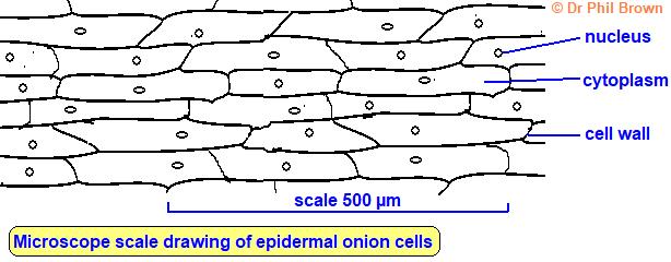
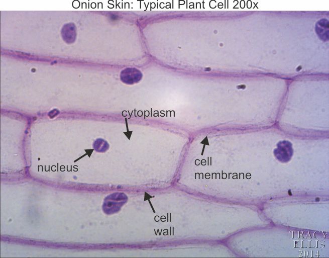
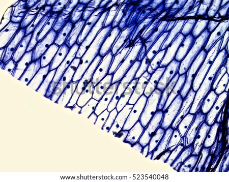
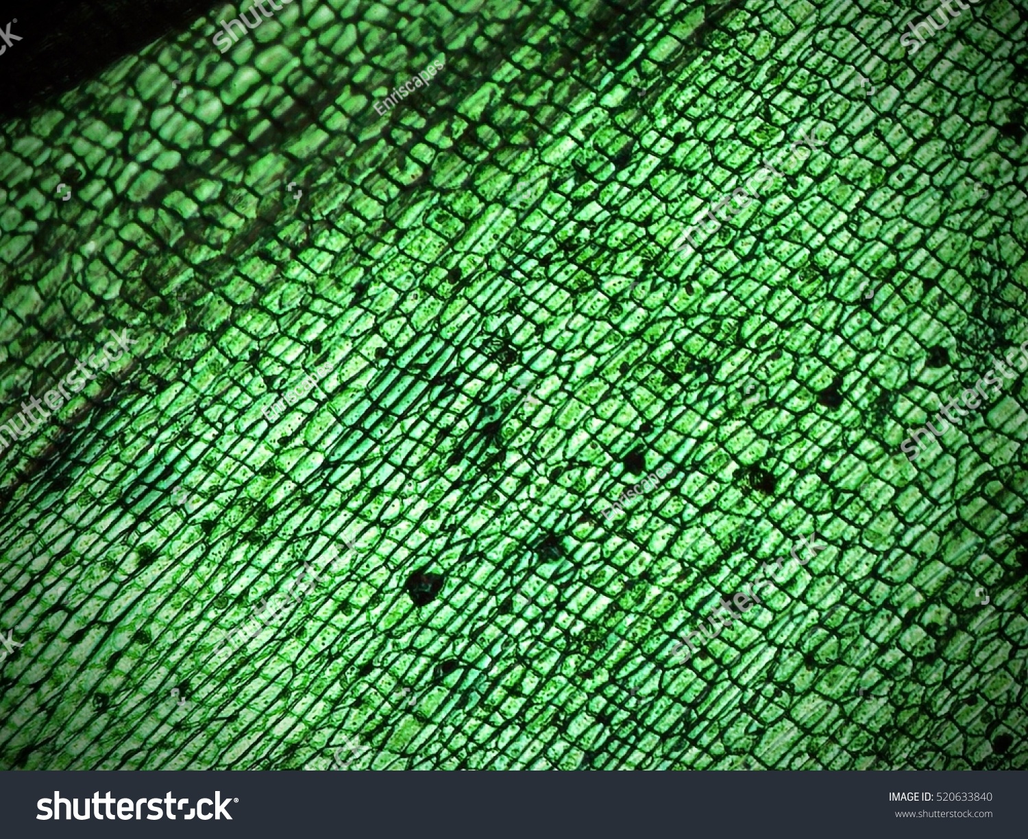
45 onion cells under microscope with labels
Cells and Reproduction - BBC Bitesize Onion cells are easy to see using a light microscope. ... A small tube placed under the skin of the upper arm. ... Five small tubes with labels and stoppers or lids Cress seeds Labels Cotton wool ... PDF Mitosis under the Microscope - Loudoun County Public Schools The chromosomes of the cells have been stained to make them easily visible. Select one cell whose chromosomes are clearly visible. 5. Sketch this cell in the appropriate box in the first row of your data chart. You are categorizing this cell in the chart according to which phase of the cell cycle/mitosis it is in. 6. Look around at the cells again.
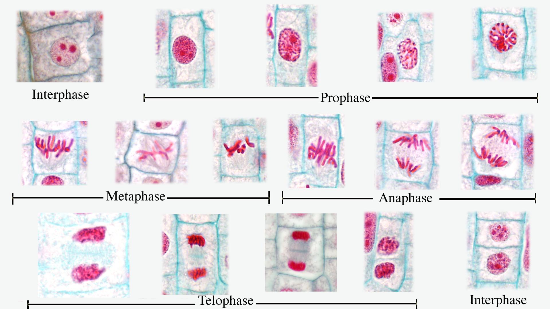
Onion Root Mitosis - Microscopy-UK Onions have larger chromosomes than most plants and stain dark. The chromosomes are easily observed through a compound light microscope. The cells pictured below are located in the apical meristem of the onion root. The apical meristem is an area of a plant where cell division takes place at a rapid rate. Phases of plant cells division:
Onion cells under microscope with labels
Collins - Concise Revision Course For CSEC Biology | PDF ... 5 Cells The cell is the basic structural and functional unit of living organisms. Some organisms are unicellular, being composed of a single cell; others are multicellular, being composed of many cells. Cells are so small that they can only be seen with a microscope and not with the naked eye. Observing Onion Cells Under The Microscope Afterwards, carefully mount the prepared and stained onion cell slide onto the microscope stage. Make sure that the cover slip is perfectly aligned with the microscope slide, and that any excess stain has been wiped off. Secure the slide on the stage using the stage clips. Under the Micrsocope: Onion Cell (100x - 400x) - YouTube In this "experiment" we will see onion cells under the microscope.For the experiment you will only need onion, dropper and the microscope (container and tool...
Onion cells under microscope with labels. Onion Cell Lab Report.docx - Onion Cell Lab Report By Onion Cell Lab Report By : Nawaf Almalki Introduction: Many things that are viewed using a microscope, particularly cells, can appear quite transparent under the microscope. The internal parts of the cells, the organelles, are so transparent that they are often difficult to see. Biologists have developed a number of stains that help them see the cells and their organelles by adding color to ... Onion Root Tip Mitosis - Stages, Experiment and Results · Cover the sample (root tip) with a coverslip and gently press the coverslip down, then examine the slide under the microscope starting with low magnification * For this experiment, a properly prepared slide should appear light pink due to the stain to almost colorless. * Unused roots can be stored in 70 percent alcohol. Results Cell Onion Labeled Under Microscope Search: Onion Cell Under Microscope Labeled. Draw & label what you observe Data: Animal Cell: Cheek Cell Plant Cell: Onion Cell Plant Cell: Elodea Cells Observation Put a drop of roughly 2% saline solution next to one edge of the cover slip and draw it under the cover slip by touching the opposite cover slip side with a paper towel, to cause This makes a root tip an excellent tissue to study ... The Cell - ScienceQuiz.net The diagram shows a group of onion cells. The parts labelled A, B and C respectively are ... The diagram shows a plant cell as seen under a microscope. Two of the ...
PDF Onion Cell Lab - somewaresinmaine.com Research Biology Onion Cell Lab page 1 of 3 Onion Cell Lab After you have completed the rest of this lab come back to this cover page DRAW & LABEL AN ONION CELL WITH ALL THE PARTS / ORGANELLES YOU OBSERVE UNDER 40X. Purpose: To observe and identify major plant cell structures and to relate the structure of the cell to its function. Materials: 1 ... Onion Cells Under a Microscope (100x-2500x) - YouTube In this video you will see onion cells under a microscope (100x-2500x) as is, without any coloring. To observe the onion cells the thin membrane is used. It... PDF Onion Cells - Investigation - Exploring Nature 5. Observe the onion tissue under the microscope at 4x, 10x and 40x with lots of light (open diaphragm). Then slowly close the diaphragm while observing the image to find the best light for seeing cellular details. 6. Draw a section of onion skin cells at 10x magnification. Then switch to 40x and draw one cell and label it. Questions: 1. Microscopy, size and magnification - Microscopy, size and ... - BBC Place cells on a microscope slide. Add a drop of water or iodine (a chemical stain). Lower a coverslip onto the onion cells using forceps or a mounted needle. This needs to be done gently to...
Plant Cell Under Microscope 40X Labeled - Powerpoint Lab Comparing ... Set up your microscope, place the onion root slide on the stage and focus on low (40x) power. 3) to draw and label a plant cell under 40x, a spider under 4x and human blood under 100x objective lens. Compare animal and plant cells and distinguish each type under the microscope. The following diagram shows cells of onion peel label class ... - Vedantu Hint: The diagrams mentioned above are the internal structure of an onion peel and human cheek cells. In order to label them, we need to understand its anatomy and know about various structures present in it. Onion peel is an example of a plant cell whereas a human cheek cell is an example of an animal cell. Complete answer: Onion Cells Microscope Stock Photos and Images - Alamy Onion cells under the microscope. Garden onion, Bulb Onion, Common Onion (Allium cepa), cell tissue of a garden onion with dyed chromosomes, light microscopy, x 120, Germany. Onion Cells under the Microscope. Onion skin cells under the microscope, horizontal field of view is about 0.61 mm. Detailed view of the cells of a red onion as seen ... Cambridge International AS and A Level Biology Coursebook ... Enter the email address you signed up with and we'll email you a reset link.
ONION CELLS VIDEO - YouTube Video shows how to make a wet mount slide to view onion cells under the microscope.
Publications – Pradeep Research Group Facile crystallization of ice I h via formaldehyde hydrate in ultrahigh vacuum under cryogenic conditions, Jyotirmoy Ghosh, Gaurav Vishwakarma, Subhadip Das, and Thalappil Pradeep, J. Phys. Chem. C, 125 (2021) 4532–4539 (DOI: 10.1021/acs.jpcc.0c10367). PDF File Supporting Information
Microscope Cell Lab: Cheek, Onion, Zebrina - SchoolWorkHelper The first lab exercise was observing animal cells, in this case, my cheek cells. The second lab exercise was observing plant cells, in this case, onion epidermis. The third lab exercise was observing chloroplasts and biological crystals, in this case, a thin section from the Zebrina plant. The first thing that was done in this lab exercise was ...
Onion Epidermal Cell Labeled Diagram - schematron.org Draw a labelled diagram of an onion epidermal cell seen under the microscope. ( 4 marks) e The onion epidermal cells are not green in colour because they lack. The epidermal cells of onions provide a protective layer against viruses and fungi that may harm the sensitive tissues.
DOC Plant and Animal Cells Microscope Lab - Hillsboro City Schools Make a drawing of one onion cell, labeling all of its parts as you observe them. (At minimum you should observe the nucleus, cell wall, and cytoplasm.) Cheek cells 1. To view cheek cells, gently scrape the inside lining of your cheek with a toothpick. DO NOT GOUGE THE INSIDE OF YOUR CHEEK! (We will observe blood cells in a future lab!!) 2.
DOC The Onion Cell Lab - chsd.us Onion tissue provides excellent cells to study under the microscope. The main cell structures are easy to see when viewed with the microscope at medium power. For example, you will observe a large circular . nucleus. in each cell, which contains the genetic material for the cell. In each nucleus, are round bodies called . nucleoli

East Central College :: programs :: Plant Mitosis Labels | Mitosis, Biology classroom, Teaching ...
OBSERVING ONION PEEL EPIDERMAL CELLS UNDER MICROSCOPE - YouTube This video is specially dedicated for my hindi subscribers. Mr Devbrat is one of them. Thank You for watching this video.This video demonstrates how to see e...
Natural Sciences Grade 9 - Grade 7-9 Workbooks The onion cells have a thick cell wall and a cell membrane. The animal cells only have a cell membrane. The onion cells have a regular shape whereas the cheek cells have a irregular shape and seem more flimsy. In the onion cells they might notice a large vacuole which might not be as visible in the cheek cells. Cheek cells do not have vacuoles ...
Onion Cells Under a Microscope - Requirements/Preparation/Observation Add a drop of iodine solution on the onion membrane (or methylene blue) Gently lay a microscopic cover slip on the membrane and press it down gently using a needle to remove air bubbles. Touch a blotting paper on one side of the slide to drain excess iodine/water solution, Place the slide on the microscope stage under low power to observe.
The Biology Project The Biology Project, an interactive online resource for learning biology developed at The University of Arizona. The Biology Project is fun, richly illustrated, and tested on 1000s of students.
Under the Micrsocope: Onion Cell (100x - 400x) - YouTube In this "experiment" we will see onion cells under the microscope.For the experiment you will only need onion, dropper and the microscope (container and tool...
Observing Onion Cells Under The Microscope Afterwards, carefully mount the prepared and stained onion cell slide onto the microscope stage. Make sure that the cover slip is perfectly aligned with the microscope slide, and that any excess stain has been wiped off. Secure the slide on the stage using the stage clips.
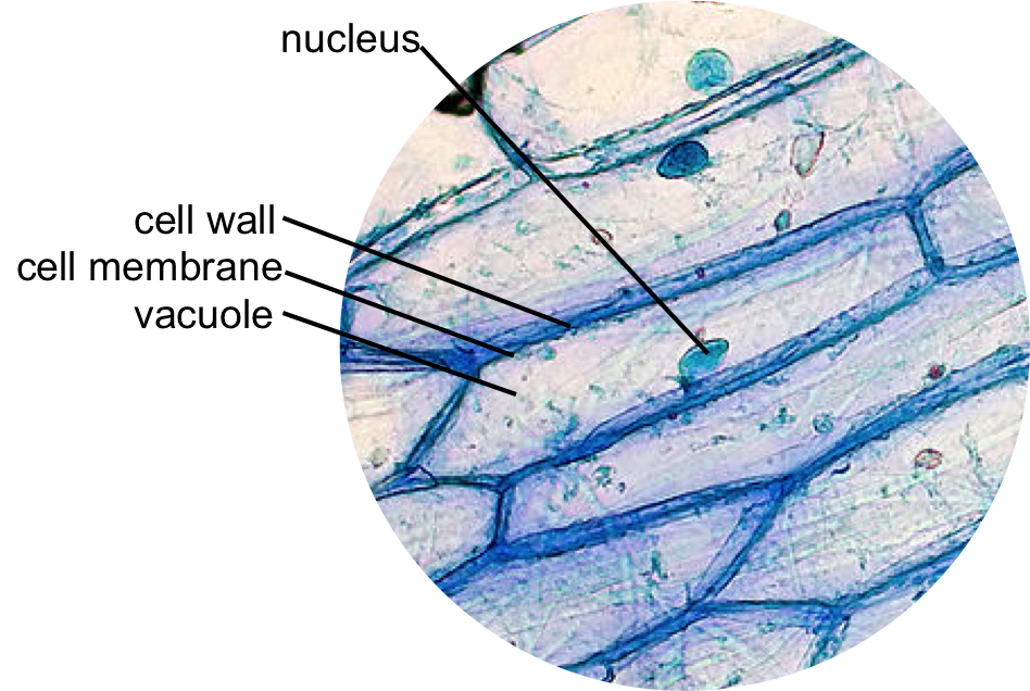
Epidermal onion cells under a microscope. Plant cells appear polygonal from the | Cell diagram ...
Collins - Concise Revision Course For CSEC Biology | PDF ... 5 Cells The cell is the basic structural and functional unit of living organisms. Some organisms are unicellular, being composed of a single cell; others are multicellular, being composed of many cells. Cells are so small that they can only be seen with a microscope and not with the naked eye.







Post a Comment for "45 onion cells under microscope with labels"