45 cat dissection pictures with labels
DOCX CAT DISSECTION - LAB REPORT - Pearland High School If you choose to take pictures, you may label the structures with tags and dissecting pins at the time of dissection if time permits or you may go back and label the pictures after pasting them in your report. The photos may be embedded into the associated sections or attached at the end of the document as an appendix. Labeled Urinary System Pictures, Images and Stock Photos Anatomy of cat with inside structure and organs scheme vector illustration. Educational veterinary and zoology study with inner system titles and location. ... System in Sagittal Section Labeled 3D Diagram of 3D computer graphic cross section of male reproductive system with labels. labeled urinary system stock pictures, royalty-free photos ...
Cat Skeleton Anatomy with Labeled Diagram - AnatomyLearner The temporomandibular joint of the cat is a synovial joint and contains an articular disc. Cat skull has a short fascial and palatal region compares to other mammals. The skull is oval elongated in shape, and has strong, highly curved zygomatic bones. There is an incomplete orbital rim in cat skull anatomy.

Cat dissection pictures with labels
Label Skeletal System Pictures, Images and Stock Photos Browse 5,663 label skeletal system stock photos and images available, or start a new search to explore more stock photos and images. Vector human anatomy, skeletal system. Female and male bodies, skeletons label skeletal system stock illustrations. Vector human anatomy, skeletal system. LifeSciTRC.org - Cat Dissection Coloring Book LifeSciTRC.org - Cat Dissection Coloring Book #R9971 Cat Dissection Coloring Book Very useful for muscles, doesn't go in superficial to deep order by page numbers, but can be easily rearranged to work that way or from anterior to posterior. — Greg Diersen, Martin Luther College Good but only shows skeletal muscles Cat Dissection - The Biology Corner Cat Dissection Cat Dissection Image Gallery 1. The largest organ is the liver, and seen embedded within the right medial lobe is the gall bladder. Not seen on this photo is the right lateral lobe which lies to the side of (and behind) the right medial lobe. The caudate lobe can also be seen by lifting the left lateral lobe. 2.
Cat dissection pictures with labels. How to Draw Animals: Cats and Their Anatomy Step 4. Let's learn how to draw the cat paw anatomy. For the front view: draw four lines ending in a "stone" shape. For the side view: draw four lines starting in an oval, and then draw a step at the end of each. The steps in the middle should be slanting. Anatomy (Dissections) - The Biology Corner Crayfish Dissection - the body cavity is exposed to reveal structures such as gills and the green gland. Crayfish Virtual Dissection - images and walk-through of crayfish dissection. Grasshopper Anatomy - examines the appendages and mouth parts. Fish Anatomy - coloring guide, fish organs and analyze fish age by scale rings. Cat Dissection Muscles Coloring Sheet | Coloring Pages Author - Unknown Date - November 09, 2016 printable coloring. Cat Dissection Muscles Coloring Sheet encouraged for you to my web site, in this particular time I am going to explain to you regarding cat dissection muscles coloring sheet. Now, this can be a very first photograph, cat dissection muscles coloring sheet : Cat Dissection Teaching Resources | Teachers Pay Teachers I made one!I have created this 32-page cat dissection manual to help guide my students through the dissection. Included in the manual are pictures of each section of the dissection. Also included are helpful hints for dissecting each section, memory hooks to help them remember specific muscles, and I have even included my dissection policies.
CAT MUSCLES - ivcc.edu CAT MUSCLES Straight from the IVCC Biology Lab to your home (without the smell)! Note: These images are best when printed using a color printer. Back and Shoulder Deep Back and Shoulder Deep Back and Shoulder with Serratus ventralis Superficial Back Neck Chest Abdominal Gluteal Dorsal Forelimb Ventral Forelimb Proximal 2021 Ultimate Veterinary Guide to Cat Anatomy with Images - VetCheck 2021 Ultimate Guide to Cat Anatomy. As the pace of veterinary advancement accelerates, even the most experienced veterinary teams are challenged to keep up with all the changes that impact their practice. ... People think and hear in pictures. Below are a selection of visual aids to help you communicate the importance of the pet's health as ... Learn How to Read a Cat X-ray | Long Beach Animal Hospital This is a labeled normal feline DV (dorsoventral) chest X-Ray of a fat cat. The top red arrow points to the aorta. The bottom red arrow points to the posterior vena cava, bringing venous blood from the back of the body to the heart . This is a normal cat lateral abdominal X-Ray L- Liver S- Stomach K- Kidneys S.I.- Small Intestines Cat Muscles (Images) Flashcards | Quizlet Cat Dissection Muscles. 31 terms. ... 41 terms. robswatski TEACHER. cat veins and arteries. 39 terms. juliadesantis1. Sets with similar terms. Cat Muscles (Images) 53 terms. meliciementor. Identify the Gender - 3rd declensions. 87 terms. Yonatan_Gut. RAD105-1 Facial Pictures. 75 terms. SithDoc89. Illustrator Tools Identification. 13 terms.
Cat Dissection Labeling Quiz - Pets and Animal Galleries Laboratory objectives, terminology, instructor commentary, dog & cat dissection videos and a dozen or so labeled cadaver images with captions are presented for each of 25 labs devoted to carnivore (dog/cat) dissection. Please try to answer all structures (or guess) before you look at the answers! Body Cavities and Organs with Labeled Diagram - AnatomyLearner While studying the gross animal anatomy, you will find different body cavities. These body cavities are the fluid-fill spaces or compartments that hold and protect the animal's internal organs. Here, in this article, I will discuss the boundary of different body cavities and organs from the animal. Animal Label Me! Printouts - EnchantedLearning.com Animal Cell Anatomy Label Me! Printout Label the animal cell diagram using the glossary of animal cell terms. Answers: Ant Anatomy: Label Me! Label the external anatomy of the ant. Answers: Australian Animals: Label Me! Label nine Australian animals. Answers: Baleen Whale Anatomy: Label Me! Label a baleen whale anatomy diagram (it's a blue ... Cat Internal Organs Photos and Premium High Res Pictures - Getty Images Browse 722 cat internal organs stock photos and images available, or start a new search to explore more stock photos and images. cross section illustration of internal anatomy of male domestic cat - cat internal organs stock illustrations. cat body vector - cat internal organs stock illustrations. checking cat's heart rate - cat internal organs ...
Cat anatomy, Dog anatomy, Cat leg
Pete The Cat Labels Teaching Resources | Teachers Pay Teachers 16. $2.50. PPTX. 6 different Pete the Cat pictures for the labels and name tags.The name tags have a dotted line to easily write students names on. Laminate the labels and use as name tags, coat rack tags, backpack tags, computer tags, use for totes, baskets, file cabinets, etc. Subjects:
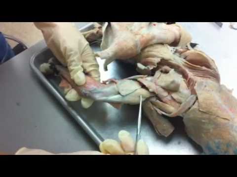
Cat Dissection Part 7 - YouTube
Feline Anatomy 101 - The Conscious Cat Your cat's mouth will be partially open when she uses this organ. This is also known as the flehmen response. Tail - it contains almost 10 percent of the cat's bones, and acts as a counterweight in helping him keep his balance. A cat's tail also communicates his mood. Understanding "tail speak" is an important part of reading feline ...
Cat dissection lab and notes chem and bio
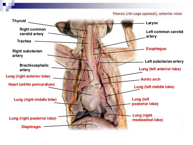
PDF Cat Anatomy Dissection Guide The cleidocervicalisis labeled clavotrapeziusin your book. This figure illustrates the position of the transversus abdominusin relation to the internaland external oblique muscles. Pelvic and Thigh Muscles - Lateral View Dorsal View - Back and Thigh Muscles Ventral View of Thigh Muscles Ventral View of Thigh and Leg Muscles The Internal Organs

Muscles Of The Chest Abdomen And Thigh (Superficial Dissection) - Conclusion moving down the ...
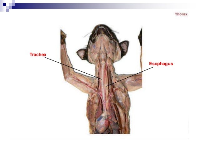
Study 132 Terms | Cat Dissection... Flashcards | Quizlet Cat Dissection Organs- Pictures STUDY Flashcards Learn Write Spell Test PLAY Match Gravity Created by Megan_Sausedo Terms in this set (132) Trachea Identify the structure at the green pointer Diaphragm Liver Identify the structure at the green pointer box. Gallbladder Identify the structure at the green pointer box. Stomach

Cat Blood Vessels Labeled - Qualitative results of our method for the blood vessels ... : during ...
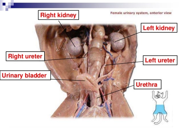
Cat anatomy - Wikipedia In the female cat, the genitalia includes the uterus, the vagina, the genital passages and teats. Together with the vulva, the vagina of the cat is involved in mating and provides a channel for newborns during parturition, or birth. The vagina is long and wide. Genital passages are the oviducts of the cat.
Cat dissection lab and notes chem and bio
Cat Anatomy | Cat Skeleton | DK Find Out Most cats have a long, slender tail made up of many small bones. The number of tail bones varies between species. For example, cats that spend a lot of time in trees, such as leopards, have longer tails to help them balance. Rib cage. The deep rib cage protects the cat's vital organs, such as the heart and lungs. Flexible spine

50 best Cat Dissection images on Pinterest | Cat anatomy, Animal anatomy and Anatomy
Digestive Cat Dissection Labeled | Virtual Anatomy Lab - ncccval Cardiovascular Sheep Heart Dissect-L. Cardiovascular Sheep Heart Disect-U. Cardiovascular Cat Dissection Labeled. Cardiovascular Cat Dissection Unlabeled. Cardiovascular Rabbit Dissection-L. Cardiovascular Rabbit Dissection -U. Respiratory. Respiratory Histology Labeled. Respiratory Histology Unlabeled.

Cat Arteries Quiz | Upper Body - (Cat manual) Lower Body - (Cat manual) | School stuff ...
PDF CAT DISSECTION A LABORATORY GUIDE - Bennington College Prepare a label for your cat with the names of your group members and the gender of your cat. 6. Follow the instructions for skinning the cat if you are dissecting skeletal muscles,or the instructions for opening the ventral body cavities if you are dissecting an organ system. FIGURE CP.1Directional terminology for the cat.

Cat dissection lab and notes chem and bio
PDF Home | Eberly College of Arts and Sciences | West Virginia University The study of cat musculature starts on p.309 of the lab manual. First sex your cat. Read over the section "Preparing the cat for muscle dissection" on p. 310. Since the pictures in your manual tend to be only close-ups, I have provided some diagrams that show the location of all the muscles so you know where they are generally located on the body.

Cat Dissection | This is a dissection of the cat, showing th… | Flickr
Cat dissection lab_labeled_images - SlideShare Cat dissection lab_labeled_images 1. Cat Dissection Muscular Labs 2. External oblique Pectroalis minor Pectoralis major Gastrocnemius Sartorius Tibialis anterior Gracilis 3.
Cat dissection lab and notes chem and bio
Cat Anatomy | Diagrams & Images of a Cats Body and Skeleton A human has 206 bones, however a cat has around 290 bones and 517 separate muscles, this makes them very agile animals, they use more than 500 muscles to leap, jump and sprint. A cat can jump over 7 times its own height. A cat has 13 ribs in its body. Take a look below at the diagram of a cats skeleton.
53 Top Pictures Cat Dissection Digestive System Labeled / Blood Vessel Cat Anatomy Dissection ...
Cat Dissection - The Biology Corner Cat Dissection Cat Dissection Image Gallery 1. The largest organ is the liver, and seen embedded within the right medial lobe is the gall bladder. Not seen on this photo is the right lateral lobe which lies to the side of (and behind) the right medial lobe. The caudate lobe can also be seen by lifting the left lateral lobe. 2.

Cat Dissection 19 | Cat Dissection | Lab Student | Flickr
LifeSciTRC.org - Cat Dissection Coloring Book LifeSciTRC.org - Cat Dissection Coloring Book #R9971 Cat Dissection Coloring Book Very useful for muscles, doesn't go in superficial to deep order by page numbers, but can be easily rearranged to work that way or from anterior to posterior. — Greg Diersen, Martin Luther College Good but only shows skeletal muscles

Cat Anatomy and Dissection with Photos – bluedoor | Top Hat - Retail
Label Skeletal System Pictures, Images and Stock Photos Browse 5,663 label skeletal system stock photos and images available, or start a new search to explore more stock photos and images. Vector human anatomy, skeletal system. Female and male bodies, skeletons label skeletal system stock illustrations. Vector human anatomy, skeletal system.
Post a Comment for "45 cat dissection pictures with labels"