38 cell diagram and labels
PDF Human Cell Diagram, Parts, Pictures, Structure and Functions One of the few cells in the human body that lacks almost all organelles are the red blood cells. The main organelles are as follows : cell membrane endoplasmic reticulum Golgi apparatus lysosomes mitochondria nucleus perioxisomes microfilaments and microtubules 2 Plant Cell: Meaning, Components, Structure, Functions & Parts - Embibe The primary function of a plant cell wall is to protect the cell against mechanical stress and to provide a definite form and structure to the cell. The cell wall consists of four layers namely: Middle lamella: Outermost cementing layer between the cells, made up of Ca and Mg pectates, absent in outer free spaces and ruptures to create ...
A Labeled Diagram of the Plant Cell and Functions of its Organelles A Labeled Diagram of the Plant Cell and Functions of its Organelles We are aware that all life stems from a single cell, and that the cell is the most basic unit of all living organisms. The cell being the smallest unit of life, is akin to a tiny room which houses several organs. Here, let's study the plant cell in detail...

Cell diagram and labels
PDF Plant Cell Diagram - Edrawsoft Plant Cell Golgi vesicles Golgi apparatus Ribosome Smooth ER(no ribosomes) Nucleolus Nucleus Rough ER(endoplasmic reticulum) Large central vacuole Amyloplast(star ch grain) Cell wall Cell membrane Chloroplast Vacuole membrane Raphide crystal Mitochondrion Druse crystal Cell Diagrams - The Biology Corner Open Google Draw and import the diagram. Then use "insert" to create text boxes where students can fill in the labels. Don't forget when assigning this to students on Google classroom to make a copy for each student. You can leave documents in an uneditable form and students can use an addon like "Kami" to annotate the document. Plant Cell - Definition, Structure, Function, Diagram & Types The plant cell wall is also involved in protecting the cell against mechanical stress and providing form and structure to the cell. It also filters the molecules passing in and out of it. The formation of the cell wall is guided by microtubules. It consists of three layers, namely, primary, secondary and the middle lamella.
Cell diagram and labels. A Well-labelled Diagram Of Animal Cell With Explanation - Byju's The animal cell diagram is widely asked in Class 10 and 12 examinations and is beneficial to understand the structure and functions of an animal. A brief explanation of the different parts of an animal cell along with a well-labelled diagram is mentioned below for reference. Also Read Different between Plant Cell and Animal Cell Animal Cells: Labelled Diagram, Definitions, and Structure The endoplasmic reticulum (s) are organelles that create a network of membranes that transport substances around the cell. They have phospholipid bilayers. There are two types of ER: the rough ER, and the smooth ER. The rough endoplasmic reticulum is rough because it has ribosomes (which is explained below) attached to it. Label Cell Parts | Plant & Animal Cell Activity | StoryboardThat Click "Start Assignment". Find diagrams of a plant and an animal cell in the Science tab. Using arrows and Textables, label each part of the cell and describe its function. Color the text boxes to group them into organelles found in only animal cells, organelles found in only plant cells, and organelles found in both cell types. Wikipedia:Featured picture candidates/Cell membrane ... Edit 3 uploaded Standard zoom boxes tend to obscure the labels, but if there is a consensus to change to those, I'll give it another try. Dhatfield ( talk ) 20:29, 16 June 2008 (UTC) [ reply ] Strong oppose There are only unsaturated tails on the phospholipids, for a reduced structure diagram of a cell membrane's lipid bilayer there should be ...
A Labelled Diagram Of Neuron with Detailed Explanations A Labelled Diagram Of Neuron with Detailed Explanations Biology Biology Article Diagram Of Neuron Diagram Of Neuron A neuron is a specialized cell, primarily involved in transmitting information through electrical and chemical signals. They are found in the brain, spinal cord and the peripheral nerves. A neuron is also known as the nerve cell. Cell: Structure and Functions (With Diagram) - Biology Discussion Eukaryotic Cells: 1. Eukaryotes are sophisticated cells with a well defined nucleus and cell organelles. 2. The cells are comparatively larger in size (10-100 μm). 3. Unicellular to multicellular in nature and evolved ~1 billion years ago. 4. The cell membrane is semipermeable and flexible. 5. These cells reproduce both asexually and sexually. Animal Cell Diagram with Label and Explanation: Cell Structure, Functions Diagram of Animal Cell Below is the diagram of the animal cell which shows the organelles present in it. The cell is covered with cytoplasm which consists of cell organelles in it. The nucleus is covered with a rough Endoplasmic Reticulum and other organelles each designed for a specific purpose. Cell Organelles- Definition, Structure, Functions, Diagram An additional non-living layer present outside the cell membrane in some cells that provides structure, protection, and filtering mechanism to the cell is the cell wall. Structure of Cell Wall. In a plant cell, the cell wall is made up of cellulose, hemicellulose, and proteins while in a fungal cell, it is composed of chitin. A cell wall is ...
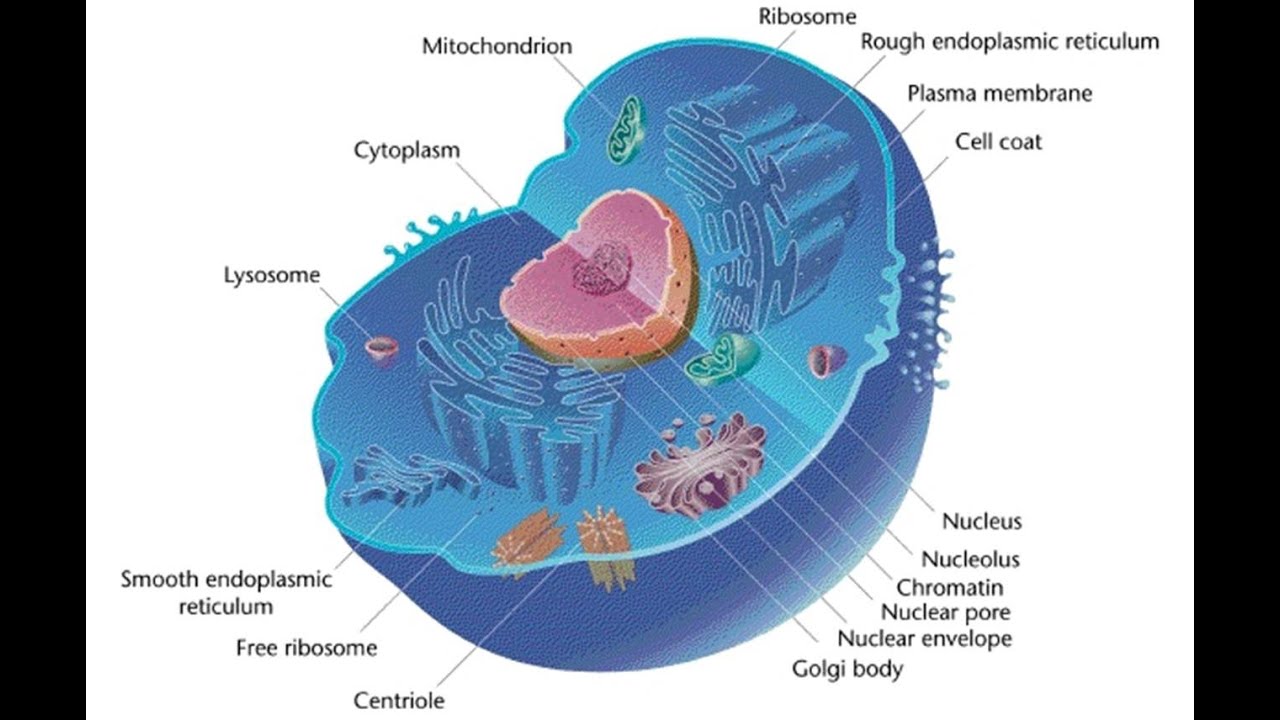
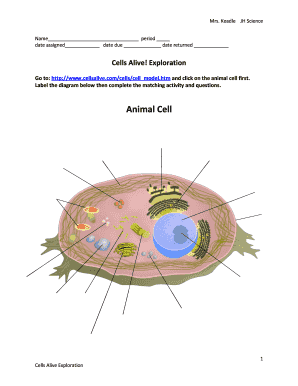
03 Label the Cell Diagram | Quizlet Cell Biology 03 Label the Cell STUDY Learn Flashcards Write Spell Test PLAY Match Gravity Created by muskopf1TEACHER Terms in this set (14) Nucleus Control center of the cell Nucleolus Ribosome synthesis Rough Endoplasmic Reticulum Protein transport Smooth Endoplasmic Reticulum Lipid synthesis Mitochondrion Cellular Respiratoin Golgi Apparatus Structure of Cell: Definition, Types, Diagram, Functions - Embibe Various kinds of cells show special differences, yet they all have some basic structural plan consisting of three essential parts: (i) cell membrane ( plasma membrane ), (ii) cytoplasm and (iii) nucleus. Apart from these three components, cells have some living parts that are called cell organelles. Human Cell Diagram, Parts, Pictures, Structure and Functions One of the few cells in the human body that lacks almost all organelles are the red blood cells. The main organelles are as follows : cell membrane endoplasmic reticulum Golgi apparatus lysosomes mitochondria nucleus perioxisomes microfilaments and microtubules Diagram of the human cell illustrating the different parts of the cell. Cell Membrane CELL MEMBRANE LABEL Diagram | Quizlet Practice labeling the parts of the cell membrane Terms in this set (6) Channel Protein hole or tunnel that particles may pass through to go in / out of cell Marker protein identifies or labels the cell Receptor protein receives information Heads part of the phospholipid that loves water (hydrophili) - points to the most outside and inside of cell
Mr. Reynolds Science Jarrell Intermediate School: May 2014
Labeled Plant Cell With Diagrams | Science Trends The parts of a plant cell include the cell wall, the cell membrane, the cytoskeleton or cytoplasm, the nucleus, the Golgi body, the mitochondria, the peroxisome's, the vacuoles, ribosomes, and the endoplasmic reticulum. Parts Of A Plant Cell The Cell Wall Let's start from the outside and work our way inwards.

This is the Cell Wall. It protects the plant cell from an...
Learn the parts of a cell with diagrams and cell quizzes Labeled cell diagram. For this exercise we'll start with an image of a cell diagram ready labeled. Study this and make sure that you're clear about which structure is found where. Cell diagram unlabeled. It's time to label the cell yourself! As you fill in the cell structure worksheet, remember the functions of each part of the cell that ...
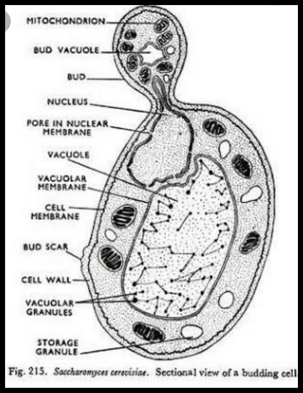
Draw a well labelled diagram of yeast - Science - Reproduction in Animals - 11997501 ...
Free Cell Diagram Software with Free Templates - EdrawMax An animal cell diagram describes a cell structure enclosed by a plasma member, and it has a nucleus with a membrane and organelles. Neuron Diagram A neuron diagram describes the three parts of a Neuron: dendrites, an axon, a cell body, or soma. Cell Membrane Diagram
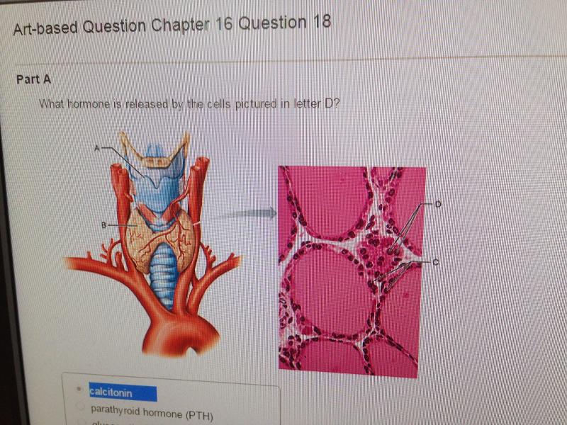
Chapter 16: Endocrine System (Mastering) Flashcards | Easy Notecards
Plant Cell - Definition, Structure, Function, Diagram & Types The plant cell wall is also involved in protecting the cell against mechanical stress and providing form and structure to the cell. It also filters the molecules passing in and out of it. The formation of the cell wall is guided by microtubules. It consists of three layers, namely, primary, secondary and the middle lamella.
Cell Diagram To Label - Pensandpieces
Cell Diagrams - The Biology Corner Open Google Draw and import the diagram. Then use "insert" to create text boxes where students can fill in the labels. Don't forget when assigning this to students on Google classroom to make a copy for each student. You can leave documents in an uneditable form and students can use an addon like "Kami" to annotate the document.
Animal cell - 3D - YouTube
PDF Plant Cell Diagram - Edrawsoft Plant Cell Golgi vesicles Golgi apparatus Ribosome Smooth ER(no ribosomes) Nucleolus Nucleus Rough ER(endoplasmic reticulum) Large central vacuole Amyloplast(star ch grain) Cell wall Cell membrane Chloroplast Vacuole membrane Raphide crystal Mitochondrion Druse crystal
Blank Animal Cell Diagram To Label

The human egg cell explained for egg donors | Altrui

LectureHub » Comparing the structures of Gram positive and negative bacteria

A Typical Cell, Labeled Stock Photo 112395266 : Shutterstock
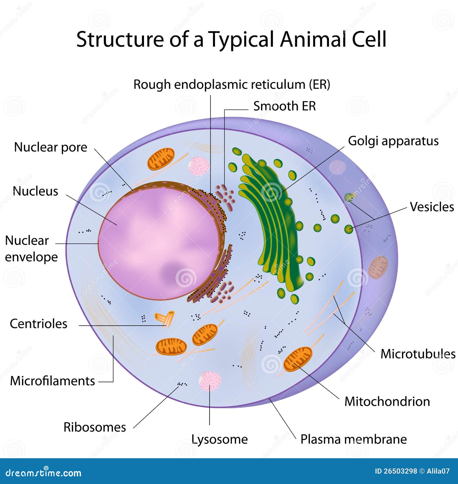
A Typical Cell, Labeled Royalty Free Stock Photos - Image: 26503298
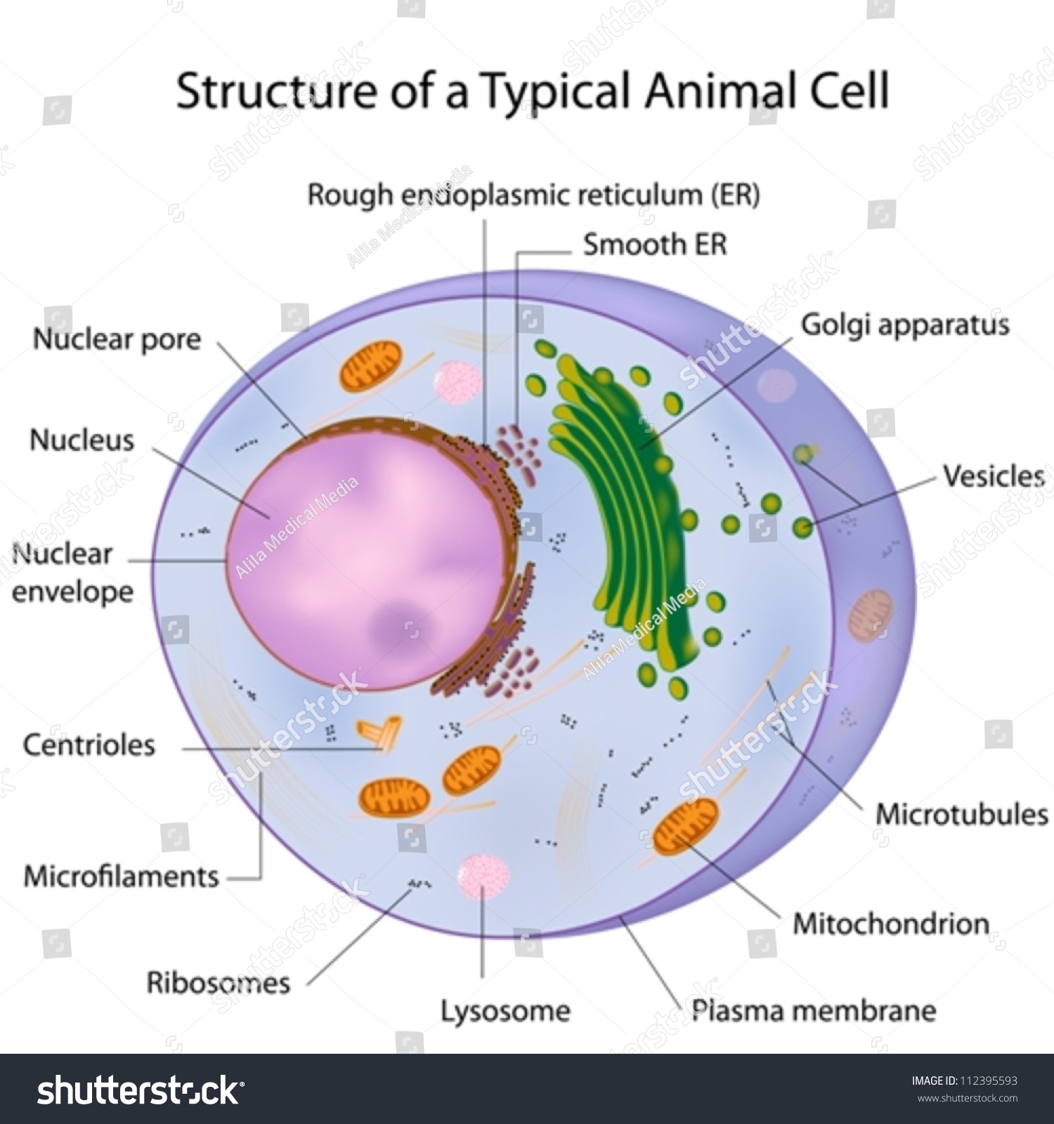
A Typical Cell, Labeled Stock Vector Illustration 112395593 : Shutterstock
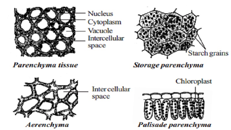
Permanent tissue: characteristics, types and functions - Online Biology Notes

Cell Diagram To Label - Pensandpieces

Animal cell structure, computer artwork - Stock Image - G450/0112 - Science Photo Library
Post a Comment for "38 cell diagram and labels"